BRITSpA Papers

BRITSpA Papers

READ HERE

BRITSpA at five

Keat A, Bennett AN, Gaffney K, Jones GT, Martindale J, Sengupta R, et al. BRITSpA at five. Rheumatology. 2020;59(4):699701. https://doi.org/10.1093/rheumatology/kez523

The British Society for Spondyloarthritis (BRITSpA) (https://britspa.co.uk/) was conceived to facilitate the sharing of information and networking among members of the various medical and scientific disciplines associated with SpA; to enhance research, diagnosis and treatment of these conditions and, importantly, to engage UK clinicians and scientists in achieving this. British clinicians and scientists have done much over the years to put SpA on the world map. The once flickering flame of ankylosing spondylitis has become an inferno of energetic and productive research. Recent developments in understanding of the disease thanks partly to the availability of MRI have even led to changed terminology for axial SpA to encompass the spectrum of inflammatory spinal disease. Combined efforts led by clinicians, academics, the pharmaceutical industry and patient organizations such as NASS have applied pressure, support and encouragement to foster better services and new knowledge. This publication introduces BRITSpA as a mechanism for mobilizing and supporting talent, ability, motivation and expertise within the UK to achieve a better future for people with SpA. In a world where substantial expertise, both pharmaceutical and academic, is being brought to bear in the field of SpA, British rheumatologists are stepping up to the mark.

READ HERE

The Use of Magnetic Resonance Imaging in Axial Spondyloarthritis: Time to Bridge the Gap Between Radiologists and Rheumatologists.

AN, Marzo-Ortega H, Kaur-Papadakis D, Rehman A. The Use of Magnetic Resonance Imaging in Axial Spondyloarthritis: Time to Bridge the Gap Between Radiologists and Rheumatologists. J Rheumatol. 2017;44(6):780-5. https://www.jrheum.org/content/44/6/780.long

Two-hundred sixty-nine radiologists from 131 acute UK National Health Service trusts/health boards responded to an online survey. Awareness of axial SpA was reported by 75% of radiologists, and awareness of definitions for positive MRI of sacroiliac joints (SIJ) and spine by 31% and 25%, respectively. These data highlight the need for better rheumatology-radiology collaboration on the identification of diagnostic axial SpA MRI lesions and support the need for a UK-based consensus on the most appropriate MRI protocols for the assessment of axial SpA (see publication below). MRI waiting times < 2 months were reported by 90% of radiologists and mean scan times were suggested at 34 minutes. However, despite internationally published consensus recommendations by The European Society of Skeletal Radiology (ESSR), the current study indicates that only 30% of radiologists scanned the whole spine and SIJ as recommended by ESSR. These modified whole-spine and SIJ protocols are cost-and time-effective, and can be performed in a standard 30-min MRI slot. Indeed, methods for using MRI in the diagnosis of axial SpA across the United Kingdom were diverse and generally did not follow the ESSR protocol. Twenty-nine radiologists (11%) used contrast as standard, 256 (91%) used T1 and short-tau inversion recovery, and 172 (64%) also used T2 sequences. Eighteen percent did not use the subchondral bone marrow edema of the SIJ to help diagnose axial SpA and 18% did not use the inflammatory vertebral corner lesions to assist diagnosis.

READ HERE

Recommendations for acquisition and interpretation of MRI of the spine and sacroiliac joints in the diagnosis of axial spondyloarthritis in the UK.

Bray TJP, Jones A, Bennett AN, Conaghan PG, Grainger A, Hodgson R, et al. Recommendations for acquisition and interpretation of MRI of the spine and sacroiliac joints in the diagnosis of axial spondyloarthritis in the UK. Rheumatology (Oxford). 2019;58(10):1831-8. https://doi.org/10.1093/rheumatology/kez173

A working group comprising nine rheumatologists and nine musculoskeletal radiologists with an interest in axial SpA was established, with support from BRITSpA. Two meetings were held. In the first meeting, research questions were formulated. In the second meeting, the results of a systematic literature review designed to inform the recommendations were reviewed. An anonymized Delphi process was used to formulate the final set of recommendations. Two overarching principles were formulated, as follows: The diagnosis of axial SpA is based on clinical, laboratory and imaging features (overarching principle 1), and patients with axial SpA can have isolated inflammation of either the sacroiliac joints or the spine (overarching principle 2). Seven recommendations addressing the use of MRI in the assessment of patients with suspected axial SpA were formulated (see table below), covering topics including recommended sequences, anatomical coverage, acquisition parameters and interpretation of active and structural MRI lesions. The level of agreement for each recommendation was very high (range 8.8–9.8). A joint rheumatology and radiology consensus on the acquisition and interpretation of MRI in axial SpA diagnosis was achieved, and a research agenda formulated. This consensus should help standardize practice around MRI and ensure a more informed, consistent approach to the diagnosis of axial SpA.

Rec1 When requesting an MRI for suspected axial SpA, imaging of both the SIJs and the spine is recommended
Rec2 T1-weighted and fat-suppressed, fluid-sensitive sequences (including STIR, fat-saturated T2 or Dixon methods) are recommended for suspected axial SpA
Rec3 
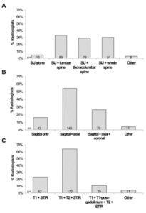
The minimum protocol when requesting an MRI for suspected axial SpA should include sagittal images of the spine with extended lateral coverage and images of the SIJs that are in an oblique coronal plane to the joint
Rec4 In the SIJs, the presence of bone marrow oedema, fatty infiltration or erosion is suggestive of the diagnosis of axial SpA. The presence of more than one of these features increases the diagnostic confidence of axial SpA
Rec5 In the spine, the presence of multiple corner inflammatory lesions and/or multiple corner fatty lesions increases the diagnostic confidence of axial SpA
Rec6 In the SIJs and/or spine the presence of characteristic new bone formation increases the diagnostic confidence of axial SpA
Rec7 The full range and combination of active and structural lesions of the SIJs and spine should be taken into account when deciding if the MRI scan is suggestive of axial SpA or not

Symptoms starting slowly

Pain in the lower back

Improves with movement

Night time waking

Early onset (under 40)